Little Known Facts About Babyecho.
Little Known Facts About Babyecho.
Blog Article
What Does Babyecho Mean?
Table of ContentsFascination About BabyechoSome Known Incorrect Statements About Babyecho Fascination About BabyechoFacts About Babyecho RevealedGet This Report on BabyechoIndicators on Babyecho You Need To KnowBabyecho - The Facts

A c-section is surgery in which your baby is born through a cut that your medical professional makes in your stomach and uterus. Whatever an ultrasound reveals, speak to your supplier about the very best take care of you and your child - fetal doppler at home. Last examined: October, 2019
During this check, they will inspect the baby is growing in the right place, whether there is greater than one baby and they will additionally check your infant's development until now. This testing is available in between 10 14 weeks of maternity and is used to examine the chances of your infant being birthed with several of these conditions.
The Ultimate Guide To Babyecho
It entails a combined test of an ultrasound scan and a blood examination. Throughout the check, the sonographer will certainly gauge the fluid at the back of the baby's neck to figure out 'nuchal clarity' - https://lwccareers.lindsey.edu/profiles/4681929-leroy-parker. They will certainly after that determine the possibility of your infant having Down's, Edwards' or Patau's disorder utilizing your age, the blood test and scan outcomes
Throughout this check, the sonographer checks for architectural and developmental abnormalities in the child. Throughout this check visit, you may be offered screenings for HIV, syphilis and hepatitis B by an expert midwife. In some situations, a third-trimester scan is advised by your midwife adhering to the results of previous tests, previous complications or existing clinical conditions.
The conventional 2D ultrasound produces level and detailed pictures which can be used to see your child's inner body organs and aid find any kind of interior issues. These black and white pictures help the sonographer determine the child's gestation, growth, heartbeat, advancement and size. Some expectant mothers pick to have a 3D ultrasound scan because they reveal more of a real-life photo of the infant.
How Babyecho can Save You Time, Stress, and Money.
3D ultrasound scans show still images of your baby's outside body instead of their insides, so you can see the form of the child's facial functions. 4D ultrasound scans resemble 3D scans but they show a relocating video clip as opposed to still images. This catches highlights and darkness much better, for that reason developing a clearer photo of the infant's face and activities.

A is discovered during this check. A lot of parents decide for this check for.
Excitement About Babyecho
Sometimes a might be called for to get and a clearer image. This is usually done and periodically a might be needed (fetal doppler). Verify that the baby's heart is present; To a lot more accurately.
Please see below. It coincides as 19-22 weeks, yet some may be or in the and it may to. Typically this is used if there are such as spina bifida or if moms and dads are keen to recognize the earlier. These scans may be done, nonetheless several of the and therefore, a is needed to This check is done usually at.
The Best Strategy To Use For Babyecho
:max_bytes(150000):strip_icc()/191127-ultrasound-trimester-pink-2000-fd089add04f8444e9d7a403933d1994f.jpg)
Additionally, the can be by by an. and is monitored by these scans. of, andare done to come to an. around the child is measured. and infant's are checked. () The method nearer the works to. Periodically, an which was previously might be.
Babyecho Things To Know Before You Get This
If, these scans might be to. (of the baby) can also be carried out. This includes, along with; This includes, along with (14-20 weeks).
A scan is essential before this test is done. If you're trying to find, set up an appointment with Dr Sankaran via her. Obstetrics & gynaecology in London.
More About Babyecho
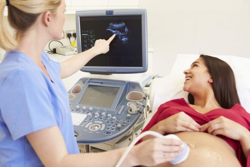
A prenatal ultrasound scan is a diagnostic strategy that utilizes high-frequency sound waves to develop a photo of your fetus. Ultrasounds might be done at various times throughout maternity for various reasons. The test can provide valuable details, aiding females and their health-care suppliers handle and care for the maternity and the fetus.
A transducer is put right into the vaginal area and rests versus the back of the vaginal canal to produce an image. A transvaginal ultrasound produces a sharper image and is frequently made use of in very early pregnancy. Ultrasound machines have to do with the size of a grocery store cart. A TV display for seeing the photos is connected to the machine (https://www.reddit.com/user/babydoppler1/).
Report this page